rs-fMRI.com
Transitioning resting-state fMRI from research theory to clinical diagnostic practice.
Module 1: The Foundation (Generalities)
Understand the physical, physiological, and mathematical principles governing functional hemodynamic brain mapping.
The BOLD Signal
Blood-Oxygen-Level-Dependent (BOLD) contrast imaging relies on the different magnetic properties of oxygenated (diamagnetic) and deoxygenated (paramagnetic) hemoglobin. It forms the foundation of all fMRI mapping.
Neurovascular Coupling
The relationship between local neural activity and subsequent changes in cerebral blood flow (CBF). This hemodynamic response is what we indirectly measure in resting-state paradigms.
Low-Frequency Fluctuations
Resting-state networks are identified by spontaneous, synchronized BOLD signal oscillations occurring at low frequencies, specifically between 0.01 and 0.1 Hz.
Historical Perspective
Trace the evolution of resting-state fMRI from Bharat Biswal's seminal 1995 discovery to the ASNR/ASFNR 2024 consensus guidelines.
Module 2: Interactive Anatomy (The 7 Yeo Networks)
Navigate the canonical functional resting-state networks and explore high-resolution clinical MPR imaging.
Network Roster
MPR Dashboard
Click over image to reveal labeling
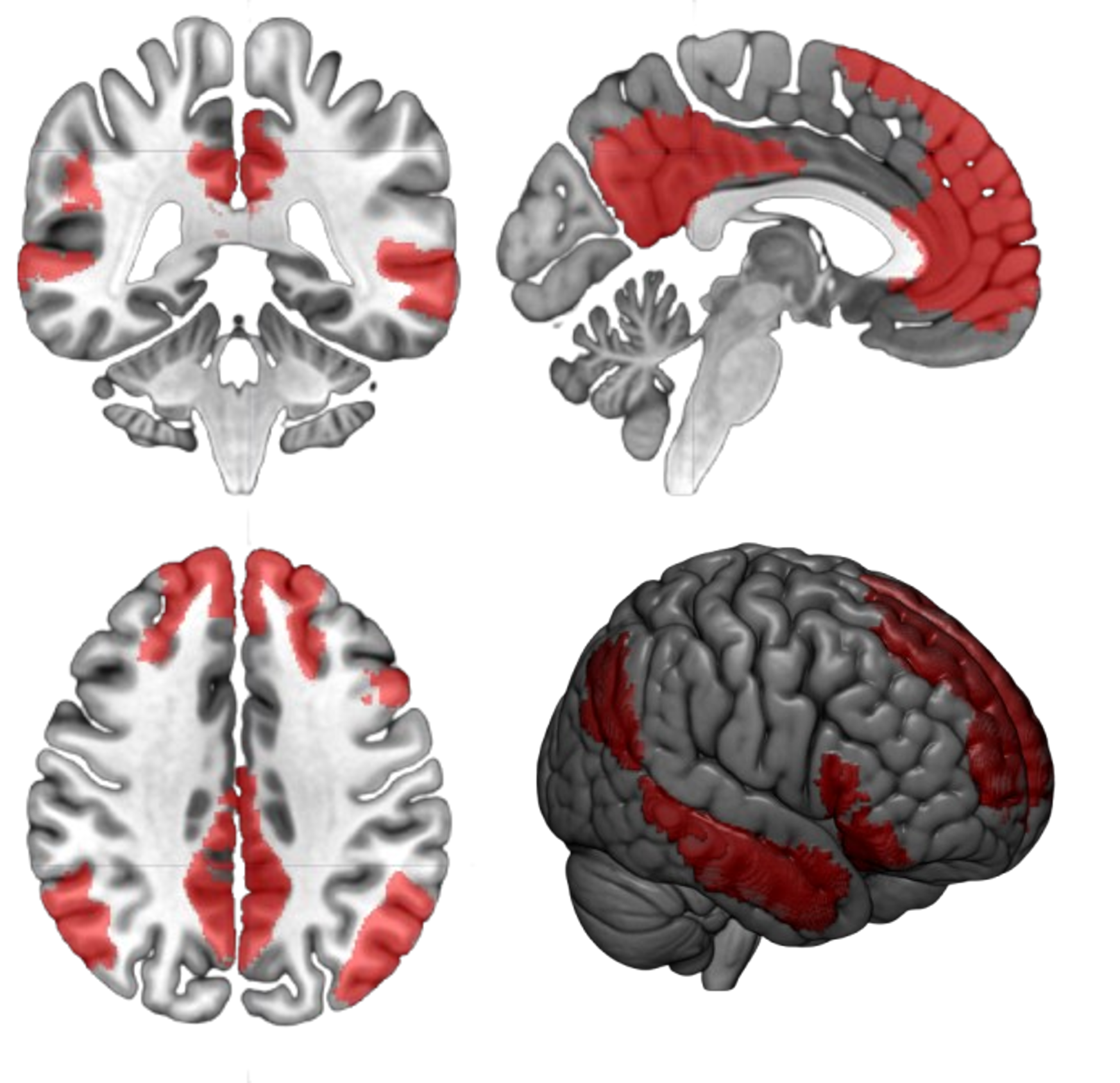
Default Mode Network
General Description
Functionally, it is the most active network during rest and introspection, linking to processes of self-referential thinking, autobiographical memory, theory of mind, and future planning, and typically showing deactivation during tasks that require external attention.
Anatomic Conventions
1. mPFC; 2. PCC/precuneus; 3. angular gyrus/inferior parietal gyrus; 4. lateral temporal cortex
Clinical Relevance
Early target for amyloid deposition in Alzheimer's disease.
Module 3: The Technical Hub (Preprocessing Workflow)
Trace the standard mathematical pipeline that prepares 4D fMRI signals for functional connectivity mapping.
Pipeline Steps
DICOM to NIfTI Conversion
This step involves critical mathematical and computational transformations applied to the 4D fMRI voxel-level signal time series.
> output shape: [64, 64, 32, 180]
> status: success
Module 4: Clinical Applications
Explore how resting-state fMRI maps translate directly into neurosurgical corridors and neuropsychiatric biomarker profiles.
Pre-Surgical Mapping
Crucial for preserving eloquent functional structures (e.g., motor strip, receptive/expressive language centers) in non-collaborative patients, pediatric populations, or those under light anesthesia where active task-based fMRI is impossible.
Network Segregation
Evaluating the breakdown of network segregation, modularity, and global efficiency using Graph Theory metrics. Serves as a vital emerging biomarker in complex neurodegenerative and psychiatric disorders.